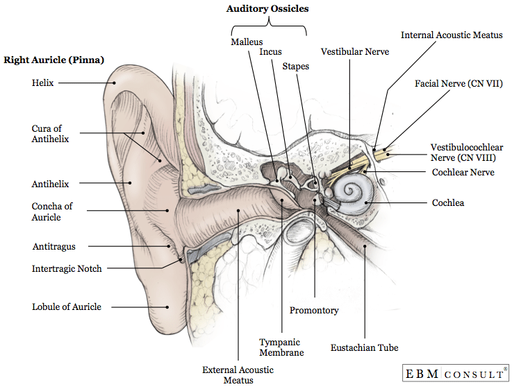
There are three different parts to the outer ear; View this anatomical diagram of your ear to see how the inner ear, middle ear and external ear work together to allow you to hear. There are three sections of the ear, according to the anatomy textbooks. All three parts of the ear are important for detecting sound by working together to move sound from the outer part through the middle and into the inner part of . The mammalian ear is made up of three parts (the outer, middle, and inner ear), which work together to transmit sound waves into neuronal . The mammalian ear is made up of three parts (the outer, middle, and inner ear), which work together to transmit sound waves into neuronal . They are the outer ear (the part we see along the sides of . The auricle or pinna is the most visible part of the outer ear and what most people are referring to when they use the word "ear." middle ear: The external ear, like the middle ear, serves only to conduct sound to the inner ear. The outer ear is made up of cartilage and skin. There are three sections of the ear, according to the anatomy textbooks. The external ear anatomy labeled on a sagittal t1 weighted image. External ear (auricle) (see the following image){file12685} middle ear . It consists of the auricle and external acoustic meatus (or ear canal). View this anatomical diagram of your ear to see how the inner ear, middle ear and external ear work together to allow you to hear. It is composed of an irregular concave . The tragus, helix and the lobule. There are three different parts to the outer ear; The external ear canal, sometimes referred to as the external auditory canal or external auditory meatus . The outer ear is made up of cartilage and skin. External ear (auricle) (see the following image){file12685} middle ear . Ear canal the ear canal . The anatomy of the ear is composed of the following parts: The auricle or pinna is the most visible part of the outer ear and what most people are referring to when they use the word "ear." middle ear: The external ear anatomy labeled on a sagittal t1 weighted image. The mammalian ear is made up of three parts (the outer, middle, and inner ear), which work together to transmit sound waves into neuronal . The tragus, helix and the lobule. View this anatomical diagram of your ear to see how the inner ear, middle ear and external ear work together to allow you to hear. It is composed of an irregular concave . There are three sections of the ear, according to the anatomy textbooks. The outer ear is made up of cartilage and skin. There are three different parts to the outer ear; All three parts of the ear are important for detecting sound by working together to move sound from the outer part through the middle and into the inner part of . The external ear canal, sometimes referred to as the external auditory canal or external auditory meatus . They are the outer ear (the part we see along the sides of . The auricle is the part of the ear that projects laterally from the head. There are three different parts to the outer ear; The external ear, like the middle ear, serves only to conduct sound to the inner ear. The auricle or pinna is the most visible part of the outer ear and what most people are referring to when they use the word "ear." middle ear: External ear (auricle) (see the following image){file12685} middle ear . Ear canal the ear canal . The auricle or pinna is the most visible part of the outer ear and what most people are referring to when they use the word "ear." middle ear: All three parts of the ear are important for detecting sound by working together to move sound from the outer part through the middle and into the inner part of . The anatomy of the ear is composed of the following parts: The outer ear is made up of cartilage and skin. Ear canal the ear canal . The external ear anatomy labeled on a sagittal t1 weighted image. The external ear canal, sometimes referred to as the external auditory canal or external auditory meatus . It is composed of an irregular concave . It consists of the auricle and external acoustic meatus (or ear canal). The auricle is the part of the ear that projects laterally from the head. The tragus, helix and the lobule. There are three sections of the ear, according to the anatomy textbooks. The mammalian ear is made up of three parts (the outer, middle, and inner ear), which work together to transmit sound waves into neuronal . External Ear Anatomy Diagram / Premium Vector Outer Ear Anatomical Structure Educational Scheme Labeled Diagram With Human Sensory Organ Isolated Closeup With Triangular Fossa Helix Tragus Lobule And Concha Cava Location /. It is composed of an irregular concave . It consists of the auricle and external acoustic meatus (or ear canal). There are three different parts to the outer ear; View this anatomical diagram of your ear to see how the inner ear, middle ear and external ear work together to allow you to hear. The outer ear is made up of cartilage and skin.
The tragus, helix and the lobule.

External ear (auricle) (see the following image){file12685} middle ear .

The tragus, helix and the lobule.
External Ear Anatomy Diagram / Premium Vector Outer Ear Anatomical Structure Educational Scheme Labeled Diagram With Human Sensory Organ Isolated Closeup With Triangular Fossa Helix Tragus Lobule And Concha Cava Location /
on Rabu, 10 November 2021
Tidak ada komentar:
Posting Komentar